October 2019
Case report- LO1 (Helderberg Hospital)Case courtesy of: Drs Dawood da Costa
1, Rena Hoffmanna1, Wiedaad Pietersen
2- Department of Medical Microbiology and Immunology, Tygerberg Hospital, Cape Town, National Health Laboratory Service
- Department of Internal Medicine, Helderberg Hospital, Somerset West, Cape Town
Written consent was obtained from the patient to publish images.
A 61-year-old male presented to the emergency department at Helderberg Hospital, Cape Town, with a 4-day history of headaches, fever, malaise, loss of appetite and a 1-day history of photophobia and confusion. Collateral history obtained in the emergency unit reported no history of vomiting, cough, night sweats, loss of weight, trauma or preceding upper respiratory tract infection symptoms.
His medical history included HIV infection diagnosed 5 years previously, and he was on a fixed dose combination of tenofovir 300mg, emtricitabine 200mg, and efavirenz 600mg antiretroviral therapy (ART). His most recent CD4 count was 330 cells/ul in May 2017, and HIV viral load (6 months before presentation, in May 2018) was less than 50 copies/ml. He had no previous TB history, no known recent household TB contact, and no recent travel history. He is a non-smoker, does not drink alcohol and adherent to his ART.
He is self-employed as a livestock farmer with pigs, goats and sheep. During the admission it was noted that he had a history of a pig bite 10 days before presentation.
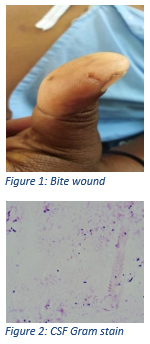
On admission he was febrile with a temperature of 38.6°C, tachycardic with a pulse of 112 bpm, normotensive with a blood pressure of 106/67 mm Hg and a fingerprick glucose test: 6.4mmol/l. He was disorientated to time, place and person with a Glasgow Coma Score of 14/15. He had evidence of a healing bite wound on his left thumb with no evidence of wound sepsis (see Figure 1). He had no evidence of anaemia, clubbing or lymphadenopathy.
Clinically, he had meningism with neck stiffness, and no focal neurological signs. He had no evidence of a skin rash, or stigmata of infective endocarditis.
His respiratory, abdominal, cardiac and neurological examination was otherwise unremarkable.
His chest x-ray was normal. Urine dipstix was normal, and bloods were drawn.
A CT brain was not performed. He was initiated on high-dose ceftriaxone 2g intravenously and admitted.